Electromagnetic Fields
An electromagnetic field is a physical field produced by electrically charged objects. These fields can produced by the local build-up of electrical charges in the atmosphere associated with thunderstorms. The earth’s own magnetic field is what causes a compass to point north. Some man-made sources of EMF’s include x-rays, electricity from every socket, TV antennas, radio stations, mobile phone stations, etc. As technology advances, EMF exposure increases.
The World Health Organization categorizes electromagnetic fields into ionizing radiation, and non-ionizing radiation. Ionizing radiation is radiation whose quanta are strong enough to break molecular bonds (ex: gamma, x-rays, radioactive materials, etc). Non-ionizing radiation is radiation whose quanta are insufficiently strong enough to break molecular bonds. Quanta are defined as the particles which carry electromagnetic waves; the higher the frequency of the EMF, the more energy and greater the “strength” of quanta.
Some possible side effects of prolonged exposure to electromagnetic fields are cancer (possible links to childhood leukemia), reduced heart rate, and interference with brain electrical activity. Interference with neural firings in the brain, and heart rate are speculated to occur during sleep. Some people have demonstrated concern relating to the EMF’s emitted from cell phones (due to the radio-frequency the cell phones give off). The Department of Energy claims “there is reason for concern,” and advice to exercise “prudent avoidance” to protect against EMF exposure.
It has been recommended by the Environmental Protection Agency that EMF’s be labeled as a class B carcinogen (defined as a probable cancer producing agent). However, the EPA recalled this claim and deemed there was not yet enough evidence to support it.
However, with this speculated threat, more research and experimentation has been performed on the effect electromagnetic fields have on the health of humans and animals. It has been realized that electric clocks have a very high magnetic field (as much as 5-10 MilliGauss up to three feet away). Sleeping with one next to your head is the equivalent of standing next to a power-line. Studies have linked high rates of brain tumors with chronic exposure (of this and similar kinds) to magnetic fields. Electric blankets and waterbed heaters create a magnetic field that penetrates 6-7 inches into the body. The “usual” and recommended ambient exposure is .5 mG. Many experts have adopted a 3.0 mG cutoff for exposure – any measure above this impedes on public safety. Dr. Nancy Wertheimer and Ed Leeper originally found the link between EMF’s and childhood leukemia, however the link is weak.
In 1974, Dr. Nancy Wertheimer was one of the first epidemiologists to study the effects of electromagnetic frequencies on biological health (namely, childhood leukemia). Pual Barodeur’s Current of Death (1989) brought the controversy to the public’s attention. However, despite Brodeur’s studies, other scientists’ data displayed no correlation between EMF exposure and health problems. A line has been drawn between varying low types of radiation. There can be either ELF or VLF frequencies (Extremely Low Frequencies, or Very Low Frequencies).
Studies performed in the field of epidemiology show the effect of EMF’s on cancer, reproduction, and neurobehavioral reactions, as well as on neurological heart and other degenerative diseases. In “positive” studies, different types of effects were recorded. No plausible and understandable mechanisms have been presented by which a carcinogenic effect could be explained. Guidelines written from these experiments “emphasize the state of scientific knowledge today does not warrant limiting exposure levels for the public and the workforce, and further data is required to confirm whether or not health hazards are present.”
During times of war, the Russians would use EMF’s to bombard the U.S. Embassy in Russia to see if there were any negative effects on behavior or thought patterns.
Some scientists are looking into a causal correlation between neurodegenerative diseases (Alzheimer’s disease, Parkinson’s disease, and Amyotrophic Lateral Sclerosis) and EMF’s. Henry Lai, Narendra P. Singh (both from the University of Washington) suggest a potential cellular mechanism for cell damage associated with EMF exposure that may help explain anomalies reported from earlier studies. They developed the Free-Radical hypothesis. This hypothesis states that extremely low frequency EMFs increase free radical activity in cells. Free radical damage can lead to cellular necrosis and apoptosis. The pair proposes that iron-rich human brain tissues (glial cells, neurons, and myelin) may be more susceptible to EMF-induced damage.
Other studies suggest that “magnetic fields created by power lines do not affect the health or reproductive capacity of farm animals or present a danger to farm fauna.” However, other studies have shown that the growth of trees which are close to transmission lines may be reduces due to the effect of corona (corona: electrical field which ionizes the surrounding atmosphere).
Caulerpa taxifolia
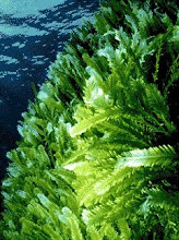
Caulerpa taxifolia is an algae species originating from the Hawaiian Islands in the United States, but has spread throughout the globe in an invasive haze. This bright green alga has a long stolon which connects individual fronds together. It attaches itself to sediments (rocks, sand, sediments, etc) with its rhizoids. The pinnules of this alga are slightly narrower and sharper than that of its almost identical cousin Caulerpa mexicana. Its frond length varies from 3-10 cm in length, with a 1-2 mm diameter. The frond length typically increases with depth. Originally, because of its bright green beauty, it was considered a novelty in aquariums. However, since its toxic invasion it has become more of a nuisance weed.
The strain originating from Hawaii is actually a harmless form. However, after cloning at the Oceanographic Museum in Monaco, the alga has been observed to have invasive properties (1989). Mutations have been observed in the nucleotides of the invasive strain as compared to the Hawaiian strain.
Many eradication methods have been attempted. Some such methods include the application of bleach to the surrounding area, removal, and covering up the algae with a tarp. However, many of these methods have not shown fruitful results. Its spread can be attributed to segmentation, boating, and aquarium usage (dumping and deporting). It was discovered off the coast of Carlsbad, California in June of 2000 – this was the first U.S. sighting of the invasive strain of Caulerpa.
Caulerpa mexicana
Caulerpa mexicana is in the empire eukaryote, kingdom plantae, subkingdom viridaeplantae, phylum chlorophyta, class bryopsidophyceae, order bryopsidales, family caulerpaceae, genus Caulerpa. It was originally named from its origin in Mexico. It is also found in Bermuda, Florida, Belize, Bahamas, Barbados, Caicos Islands, Cayman Islands, Cuba, Hispaniola, Jamaica, Lesser Antilles, Puerto Rico, Tobago, Virgin Islands, Brazil, Columbia, Guyana, Venezuela, and Madagascar. Nucleotide sequences in both Caulerpa mexicana, and Caulerpa taxifolia have shown genetic similarities between the two cousins. This explains the visible similarities between the two algae.
Caulerpa mexicana can be found on sandy bottoms (often on mounds of sand and sometimes on hard surfaces). Seasonally abundant, and can cover large areas when in “bloom.” Similar to Caulerpa taxifolia, Caulerpa mexicana is a feathery structure with fronds which are 5-6 cm long. The central frond is flat, while the pinnules are short, flat and have slightly rounded tips. These immerge from a creeping stolon that has small rhizoids to attach the algae to hard sediments. The color is usually some shade of green.
Photosynthesis
All forms of Caulerpa photosynthesize. Photosynthesis is the process of converting light energy to chemical energy and storing it in bonds of sugar. The materials necessary for a plant to photosynthesize are as follows: CO2, light energy, and H2O. The process takes place in the chloroplasts using chlorophyll. Chlorophyll is a green pigment involved in photosynthesis. Some other pigments involved with photosynthesis are carotenoids (contains carotene. Made of two small six carbon rings connected in a chain of carbon atoms. Called “accessory pigments”), and phycrobilins (found in cytoplasm or stroma of chloroplasts. Occur in cyanobacteria and rhodophyta.
Chlorophyll contains a pophyrin ring. The stability of this molecule allows electrons to migrate. This increases the rings potential to gain or lose electrons (this is how chlorophyll captures light. Chlorophyll “a” passes its energized electrons on to molecules to make sugars.
The chloroplasts are made up of outer and inner membranes, intermembrane space, stroma, and thylakoids stacked in grana. Chlorophyll is built into membranes of the thylakoid. The chemical equation for photosynthesis is 6CO2 + 6H2O + light energy -> C6h12O6 + 6O2. The C6H12O6 is glucose. One process in photosynthesis is the light reactions. This is where the plant converts light energy into chemical energy in the thylakoid membrane. Chlorophyll and other pigments absorb various wavelengths of light and pass their energy to the central chlorophyll molecule. Chlorophyll consists of several fused rings of carbon and nitrogen with a magnesium ion in the center.
Another portion of photosynthesis is the Calvin cycle which spends ATP and consumes NADPH2 to produce glucose. Phase One of the Calvin cycle is carbon fixation.
In carbon fixation, RuBP carboxylase catalyzes; CO2 is incorporated into a five-carbon sugar named ribulose biphosphate; a 6 carbon intermediate is produced, then splits in half to form 2 molecules of 3-phosphoglycerate.
Phase Two is reduction. This is where ATP converts the 3-phosphoglycerate into glyceraldehyde 3-phosphate. Then, in Phase Three is where the product of Phase two back into RuBP and put again through the Calvin Cycle for later use.
Another cycle in photosynthesis is the carbon cycle. In the carbon cycle, the absorbed CO2 and sunlight are used to create fuel-glucose and other sugar used for further growth and building of the plant structure. The plants go through respiration where it releases energy contained in sugars to use it in the plant metabolism. It also renders the carbohydrates “fuel” back to CO2. Decomposition is respiration that consumes organic matter mostly by bacteria or fungi. During times when photosynthesis exceeds respiration, organic matter is slowly built up and forms coal and oil deposits (over millions of years).
Chlorophyll
Chlorophyll is a green photosynthetic pigment found in all photosynthesizing organisms. Chlorophyll is capable of channeling the energy of sunlight into chemical energy through the process of photosynthesis. It assists in the transfer of electrons from water to carbon dioxide. Electrons within chlorophyll are excited from a lower energy state to a higher energy state as the chlorophyll absorbs light. Due to its higher energy state, the electron is more readily transferred during photosynthesis. This transfer begins the electron-transfer process which ultimately ends with carbon dioxide gaining the electron from the chlorophyll. Chlorophyll is at the center of the photosynthetic oxidation-reduction reaction between carbon dioxide and water.
In plants, chlorophyll is associated with specific proteins, for example, chlorophyll-a binding proteins are referred to as CP I, CP 47, and CP 43. With improving biochemical techniques for use on the membrane systems there has been an increase in the success of isolation and characterization of these proteins. Chlorophyll is also essentially made of two parts – a substituted porphyrin ring and phytol (long carbon chain).
Originally it was assumed that chlorophyll was a single compound. However, experiments in 1864 showed by spectroscopy that chlorophyll was a mixture. Stokes showed that if dried leaver are powdered and digested with ethanol ‘crystalline’ chlorophyll is obtained. However, when ether or aqueous acetone is used instead of ethanol, the resultant is ‘amorphous’ chlorophyll. In 1912, Wilstatter et al. showed that chlorophyll was a mixture of two compounds, chlorophyll-a and chlorophyll-b. In natural chlorophyll there is a ration of 3:1 (of a to b) of the two components.
Caulerpa (w/Diver)

Level of infestation in a given area.


